:main_page.jpg
About
The Brain Image Analysis Unit leverages the latest advancements in machine learning, specifically deep learning, to enhance the processing and analysis of brain imaging data. Our unit specializes in processing diverse brain imaging data sources, including two-photon microscopy, bright-field microscopy images, and MRI scans. We collaborate closely with experts from various scientific disciplines, including computer science, neuroscience, and medicine, to advance the state-of-the-art in brain imaging analysis. As a member of the Brain/MINDS 2.0 project, our unit is dedicated to uncovering new insights into the intricacies of primate brain structure through the analysis of high-resolution brain imaging data.
Members

Henrik Skibbe
Unit Leader

Itsuko Ishii
Technical Staff

Peerawat Pannattee
Postdoctoral researcher

Méghane Decroocq
Visiting Scientist
(Assistant Professor,
TOKYO INTERNATIONAL UNIVERSITY)

Febrian Rachmadi
Visiting Scientist
Faculty of Computer Science, Universitas Indonesia

Michał Byra
Visiting Scientist
(Assistant Professor,
Polish Academy of Sciense,
Warsaw)

Charissa Poon
Visiting Scientist
Tools and Data
- Our GitHub page: https://github.com/BrainImageAnalysis.
- The BMCR Resources moved to a new place
News
- 2026
- A MIDL short-paper has been accepted. Congratulations to Peerawat! 🎉
- A MIDL short-paper has been accepted. Congratulations to Meghane! 🎉
- 2025
- Peerawat has joined us as Postdoctoral researcher
- A paper has been accepted for presenting at the MICCAI 2025 Workshop on Shape in Medical Imaging! 🎉 Congratulations to Meghane! 🎉
- Our Nature Scientific Reports paper is online! 🎉 Congratulations to Febrian! 🎉
- 2024
- The BMCR related content has been moved to a new homepage: https://bmcr.brainminds.jp/
- A MIDL short-paper has been accepted. Congratulations to Charissa! 🎉
- A MIDL paper has been accepted. Congratulations to Meghane! 🎉
- Our paper about instance-level loss functions is online. Congratulations to Febrian! 🎉
- 2023
- An image generated in our group made it onto the front page of RIKEN Research Winter 2023
- Méghane Decroocq has joined us as a JSPS postdoctoral fellow.
- Our Nature Scientific Reports paper is online! 🎉
- Our contribution to the Brain/MINDS Nature Advertisement feature is online
- A MICCAI paper has been accepted. Congratulations to Michal! 🎉
- Our PLoS Biology paper is online! 🎉
- An MIDL paper has been accepted. Congratulations to Febrian! 🎉
- An ISBI paper has been accepted. Congratulations to Charissa! 🎉
- 2022
- Binbin Xu has joined us as a Visiting Researcher.
- Michal Byra has joined us as a JSPS postdoctoral fellow.
- Our new website is online! 🎉
- Charissa's Kakenhi proposal has been accepted! 🎉😻
- Michal Byra has joined us as a visiting researcher.
- Matthias Schlachter has joined us as a Riken Special Postdoctoral Researcher.
- 2021
- The Best Paper Award for the 2021 PRIME workshop was awarded to Febrian! 🎉
- Febrian became a Riken Special Postdoctoral Researcher.
- Febrian's Kakenhi proposal has been accepted! 🎉
- Charissa Poon joined us as a postdoctoral researcher.
- Yasutaka Odo has started working with us as a student trainee.
- マーモセットの遺伝子発現データベースを公開 (press release)
- 2020
- Marco Reisert has joined us as a visiting researcher.
- Japan’s big brain project: advances light up marmoset brains (press release)
- Febrian Rachmadi joined us as a research scientist.
Our Research On Cover Pages

Our brain images appeared on a cover of the MIDL (Medical Imaging with Deep Learning) 2021 conference magazine.
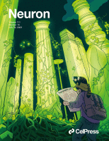
Our research inspired a Neuron cover. Artwork by Natsuko Miyazaki (Space-Time Inc.).

Our brain connectivity image was used in a cover design of the RIKEN RESEARCH magazine.

One of our images showing brain connectivity was used for the design of a book cover.

The cover page features a neuron image created by Kimura et al. as part of a workflow demonstration for software designed for terminal detection.
Alumni and previous guests
- Gaganpreet Jhajj: Summer intern in 2025
- Matthias Schlachter : Special Postdoctoral Researcher 2022-2024
- Jeanne Salle: Summer intern in 2023
- Binbin Xu : (Associate Professor, MT Mines Ales, Biomedical Data Science) Visiting Scientist 2022
- Yasutaka Odo : Student trainee and research part-time worker from 2021-2022.
- Faegheh Yeganli : Research scientist in 2020.
Publications
( 📚published manuscript, conference paper or short paper, 📢abstract, 📝 preprint)
2026
-
- M. Decroocq, K. Onishi, T. Shimogori, H. Skibbe
- In MIDL, 2026
-
- P Pannattee, A Watakabe, T Shimogori, H Skibbe
- In MIDL, 2026
-
- T. Yamamori, A. Watakabe, H. Skibbe
- In Frontiers in Neuroanatomy, 2026
-
- V. A Coenen, A. Rau, A. Watakabe, H. Skibbe, T. Yamamori, T. E Schläpfer, M. Czornik, D. Meyer-Doll, D. Endres, J. C. Baldermann, H. Urbach, M. D Döbrössy, B. EA Sajonz, M. Reisert
- In Frontiers in Research Square, 2026
-
- H. Skibbe, M. Byra, A. Watakabe, T. Yamamori, M. Reisert
- In Scientific Reports, 2026
2025
-
- K. Dewa, K. Kaseda, A. Kuwahara, H. Kubotera, A. Yamasaki, N. Awata, A. Komori, M. A. Holtz, A. Kasai, H. Skibbe, N. Takata, T. Yokoyama, M. Tsuda, G. Numata, S. Nakamura, E. Takimoto, M. Sakamoto, M. Ito, T. Masuda & J. Nagai
- In Nature, 2025
-
- K Fujimoto, S. Ishii, R. Gong, K. Nakae, N. Ichinohe, and H. Skibbe
- Accepted for presenting at ICONIP 2025
-
- M. Decroocq, C. Poon, M. Schlachter , H. Skibbe
- Accepted for presenting at MICCAI 2025 Workshop on Shape in Medical Imaging, 2025
-
- M. Byra, H. Skibbe
- IEEE/CVF Winter Conference on Applications of Computer Vision (WACV), 2025
-
- M. F. Rachmadi, M. del C. Valdés-Hernández, S. Makin, J. Wardlaw , H. Skibbe
- In Scientific Reports, 2025
-
- A. Watakabe, T, Tani, H. Abe, H. Skibbe, N. Ichinohe, T. Yamamori
- Journal of Visualized Experiments (JoVE), 2025
2024
-
- A. Watakabe, T, Tani, H. Abe, H. Skibbe, N. Ichinohe, T. Yamamori
- In bioRxiv, 2024
-
- H. Ju, H. Skibbe, M. Fukui, S. H. Yoshimura, H. Naoki
- iScience, 2024
-
- C. Poon, M. Byra, T. Shimogori, H. Skibbe
- MIDL, 2024, accepted
-
- M. F. Rachmadi, M. Byra, H. Skibbe
- In Computers in Biology and Medicine, 2024
-
- M. Decroocq, B. XU, K. L. Thompson-Peer, A. Moore, H. Skibbe
- MIDL, 2024, accepted
-
- M. F. Rachmadi, P. Kreshanti, M. I. Anggraeni,V. Tania, R. E. Yunus, H. Skibbe
- In TechRxiv, 2024
-
- H. Ju, H. Skibbe, M. Fukui, S. H. Yoshimura, H. Naoki
- In bioRxiv, Cold Spring Harbor Laboratory, 2024
-
- C. Poon, M. Byra, M. F. Rachmadi, M. Schlachter, M. Decroocq, B. Xu, B. Fulcher, T. Shimogori, H. Skibbe
- Neuroscience, 2024, (Fukuoka, Japan)
-
- H. Tsukada, K. Nakae, J. Hata, H. T. Hamada, K. Tokuda, C. E. Gutierrez, H. Skibbe, C. Poon, A. Woodward, S. Ishii, T. Shimogori, H. Okano, K. Doya
- Neuroscience, 2024, (Fukuoka, Japan)
2023
-
- M. Kimura, J. Tann, O. Wilkes, F. Xu, H. Skibbe, A. W. Moore
- Cold Spring Harbor Protocols, 2023
-
- Tann JY, Xu F, Kimura M, Wilkes OR, Yoong LF, Skibbe H, Moore AW.
- Cold Spring Harbor Protocols, 2023
-
- Y. Shima, H. Skibbe, Y. Sasagawa, N. Fujimori, Y. Iwayama, A. Isomura-Matoba, M. Yano, T. Ichikawa, I. Nikaido, N. Hattori, T. Kato
- In Cell reports, 2023
-
- M. Byra, C. Poon, M. F. Rachmadi, M. Schlachter, H. Skibbe
- In Scientific reports, Nature Publishing Group, 2023.
-
- M. Byra, C. Poon, T. Shimogori, H. Skibbe
- MICCAI, 2023
-
- H. Skibbe, M.F. Rachmadi, K. Nakae, C. E. Gutierrez, J. Hata, H. Tsukada, C. Poon, K. Doya, P. Majka, M. G. P. Rosa, M. Schlachter, H. Okano, T. Yamamori, S. Ishii, M. Reisert, A. Watakabe.
- In PLoS Biology, 2023
-
- A. Watakabe, H. Skibbe, K. Nakae, H. Abe, N. Ichinohe, M. F. Rachmadi, J. Wang, M. Takaji, H. Mizukami, A. Woodward, R. Gong, J. Hata, D. C. Van Essen, H. Okano, S. Ishii, T. Yamamori
- In Neuron, 2023
-
- M.F. Rachmadi, C. Poon, H. Skibbe
- MIDL, 2023
-
- V. A. Coenen, A. Watakabe, H. Skibbe, T. Yamamori, M. D. Döbrössy, B. E.A. Sajonz, P. C. Reinacher, M. Reisert
- In Brain Stimulation, 2023
-
- C. Poon, M.F. Rachmadi, M. Byra, M. Schlachter, B. Xu, T. Shimogori, H. Skibbe
- In IEEE International Symposium on Biomedical Imaging, to appear.
-
- C. Poon, Teikari P., M.F. Rachmadi, H. Skibbe, K. Hynynen
- In Scientific Data, 2023
-
- M, Byra, M. F. Rachmadi, H. Skibbe
- In arXiv, 2023
-
- H. Skibbe, M. Byra, A. Watakabe, T. Yamamori, M. Reisert
- In arXiv, 2023
- 📢 Deep Learning-Based Multi-Modal Image Processing Using an Interactive Web-Platform: Segmenting and Identifying Neurons in Drosophila
- M. Schlachter, M. Someya, H. Kazama, H. Skibbe
- Neuroscience, 2023. (Sendai, Japan)
- 📢 An automated pipeline to create a gene expression atlas in the marmoset brain
- C. Poon, M. F. Rachmadi, M. Byra, M. Schlachter, B. Xu, T. Shimogori, H. Skibbe
- Neuroscience, 2023. (Sendai, Japan)
2022
- 📚Where Position Matters - Deep learning driven normalization and co-registration of computed tomography in the postoperative analysis of Deep Brain Stimulation
- M. Reisert, B. Sajonz, P. Reinacher, M. Russe, E. Kellner, H. Skibbe, V. A. Coenen
- In Neuromodulation: Technology at the Neural Interface, to appear
-
- C. E. Gutierres, H. Skibbe, H. Musset, K. Doya.
- Frontiers in Neuroinformatics (to appear), 2022.
-
- M.F. Rachmadi, M.d.C. Valdés-Hernández, S. Makin, J. Wardlaw, H. Skibbe
- In bioRxiv, Cold Spring Harbor Laboratory, 2022.
-
- J. Hata, K. Nakae, H. Tsukada, A. Woodward, Y. Haga, M. Iida, A. Uematsu, F. Seki, N. Ichinohe, R. Gong, T. Kaneko, D. Yoshimaru, A. Watakabe, H. Abe, T. Tani, H. Skibbe, M. Maeda, F. Papazian, K. Hagiya, N. Kishi, T. Shimogori, T. Yamamori, H. James Okano, H. Okano
- In bioRxiv, Cold Spring Harbor Laboratory, 2022.
-
- H. Skibbe, M.F. Rachmadi, K. Nakae, C. E. Gutierrez, J. Hata, H. Tsukada, C. Poon, K. Doya, P. Majka, M. G. P. Rosa, H. Okano, T. Yamamori, S. Ishii, M. Reisert, A. Watakabe.
- In bioRxiv, Cold Spring Harbor Laboratory, 2022.
-
- Shima, Y., Skibbe, H, Sasagawa, Y., Fujimori, N., Nikaido, I., Hattori, N. and Kato, T.
- In bioRxiv, Cold Spring Harbor Laboratory, 2022.
- 📢 Semi-supervised semantic segmentation of in situ hybridization gene expression in the marmoset brain
- C. Poon, M.F. Rachmadi, M. Byra, T. Shimogori, and H. Skibbe
- Society for Neuroscience, 2022. (California, USA)
- 📢 Semi-supervised contrastive learning for semantic segmentation of in situ hybridization gene expression in the marmoset brain
- C. Poon, M.F. Rachmadi, M. Byra, T. Shimogori, and H. Skibbe
- International Symposium on Artificial Intelligence and Brain Science, 2022. (Okinawa, Japan)
- 📢 Development of a Data-driven Prediction Model for the Evolution of White Matter Hyperintensities using Deep Learning: Progress and Challenges
- M.F. Rachmadi, M.d.C. Valdés-Hernández, S. Makin, J.M. Wardlaw, T. Komura, and H. Skibbe
- Neuroscience, 2022. (Okinawa, Japan)
- 📢 Development of a Data-driven Prediction Model for the Evolution of White Matter Hyperintensities using Deep Learning: Progress and Challenges
- M.F. Rachmadi, M.d.C. Valdés-Hernández, S. Makin, J.M. Wardlaw, T. Komura, and H. Skibbe
- I nternational Symposium on Artificial Intelligence and Brain Science, 2022. (Okinawa, Japan)
- 📢 Semi-supervised contrastive learning for semantic segmentation of in situ hybridization gene expression in the marmoset brain
- C. Poon, M.F. Rachmadi, M. Byra, T. Shimogori, and H. Skibbe
- Neuroscience, 2022. (Okinawa, Japan)
2021
-
- Rachmadi, M. F., Valdés-Hernández, M. del C, Maulana, R., Wardlaw, J., Makin, S. and Skibbe, H.
- In International Workshop on PRedictive Intelligence In MEdicine, 2021.
-
- Kita, Y., Nishibe, H., Wang, Y., Hashikawa, T., Kikuchi, S. S., Mami, U,. Yoshida, A. C., Yoshida, C., Kawase, T., Ishii, S., Skibbe, H. and Shimogori, T.
- In Proceedings of the National Academy of Sciences, National Acad Sciences, volume 118, 2021.
-
- Skibbe, H., Watakabe, A., Rachmadi, F., Gutierrez, C.E, Nakae,K. and Yamamori, T.
- In Medical Imaging with Deep Learning 2021, 2021.
-
- Watakabe, A., Skibbe, H. Nakae, K., Abe, H., Ichinohe, N., Wang, J., Takaji, M., Mizukami, H., Woodward, A., Gong, R., Hata, J., Okano, H., Ishii, S. and Yamamori, T.
- In bioRxiv, Cold Spring Harbor Laboratory, 2021.
2020
-
- Gutierrez, Carlos Enrique, Skibbe, Henrik, Nakae, Ken, Tsukada, Hiromichi, Lienard, Jean, Watakabe, Akiya, Hata, Junichi, Reisert, Marco, Woodward, Alexander, Yamaguchi, Yoko, Yamamori, Tetsuo, Okano, Hideyuki, Ishii,Shin and Doya, Kenji
- In Scientific reports, Nature Publishing Group, volume 10, 2020.
2019
- 📢 Mapping connectivity of the marmoset prefrontal cortex
- A. Watakabe, H. Skibbe, K. Nakae, J. Wang, M. Takaji, H. Mizukami, A. Woodward, R. Gong, Y. Yamaguchi, J. Hata, H. Okano, S. Ishii and T. Yamamori
- Neuroscience, 2019. (Niigata, Japan)
- 📢 Fully automated data processing for mapping connectivity of the marmoset prefrontal cortex
- H. Skibbe, A. Watakabe, K. Nakae, C. E. Gutierrez, A. Woodward, H. Tsukada, R. Gong, J. Hata, K. Doya, H. Okano, T. Yamamori and S. Ishii
- Neuroscience, 2019. (Niigata, Japan)
- 📢 A fully automated, AI-driven pipeline for the determination of the marmoset brain connectivity based on tracer data obtained with the TissueCyte microscope
- H Skibbe, A. Watakabe, K. Nakae, C. E. Gutierrez, A. Woodward, H. Tsukada, R. Gong, J. Hata, H. Okano, T. Yamamori and S. Ishii
- International Symposium of Brain/MINDS ISBM, 2019. (Tokyo, Japan)
- 📢 Macro-scale connectome by diffusion MRI of exvivo marmoset brain with a pipeline of global fiber reconstruction
- K. Nakae, J. Hata, H. Skibbe, A. Woodward, C. E. Gutierrez, H. Tsukada, G. Rui, H. Okano and S. Ishii
- International Symposium of Brain/MINDS ISBM, 2019. (Tokyo, Japan)
- 📢 Multi-objective Parameter Optimization of DWI-based Global Fiber Tracking with Neuronal Tracer Signal as a Reference
- C. Enrique Gutierrez, H. Skibbe, K. Nakae, J. Liénard, A. Woodward, A. Watakabe, H. Tsukada, J. Hata, H. Okano, T. Yamamori, Y. Yamaguchi, S. Ishii and K. Doya
- International Symposium of Brain/MINDS ISBM, 2019. (Tokyo, Japan)